


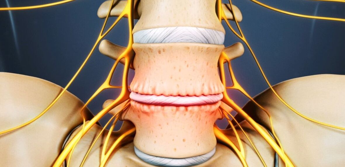
Osteochondrosis is a chronic degenerative-dystrophic disease that is caused by a number of quite different factors. Pathological lesions initially occur in the nucleus pulposus (the inner contents of the intervertebral disc) and then spread to the fibrous ring (the outer shell of the disc) and other elements of the spinal movement segment (SDS). This may be due to the body’s natural aging process, or it may be due to injuries, increased spinal strain, and other causes. In any case, osteochondrosis is only the first stage in the destruction of the intervertebral disc, and if left untreated, protrusions and hernias develop that often require surgical removal.
The intervertebral disc is a cartilage formation that separates the vertebral bodies and acts as a shock absorber.
Lumbar osteochondrosis: what is it
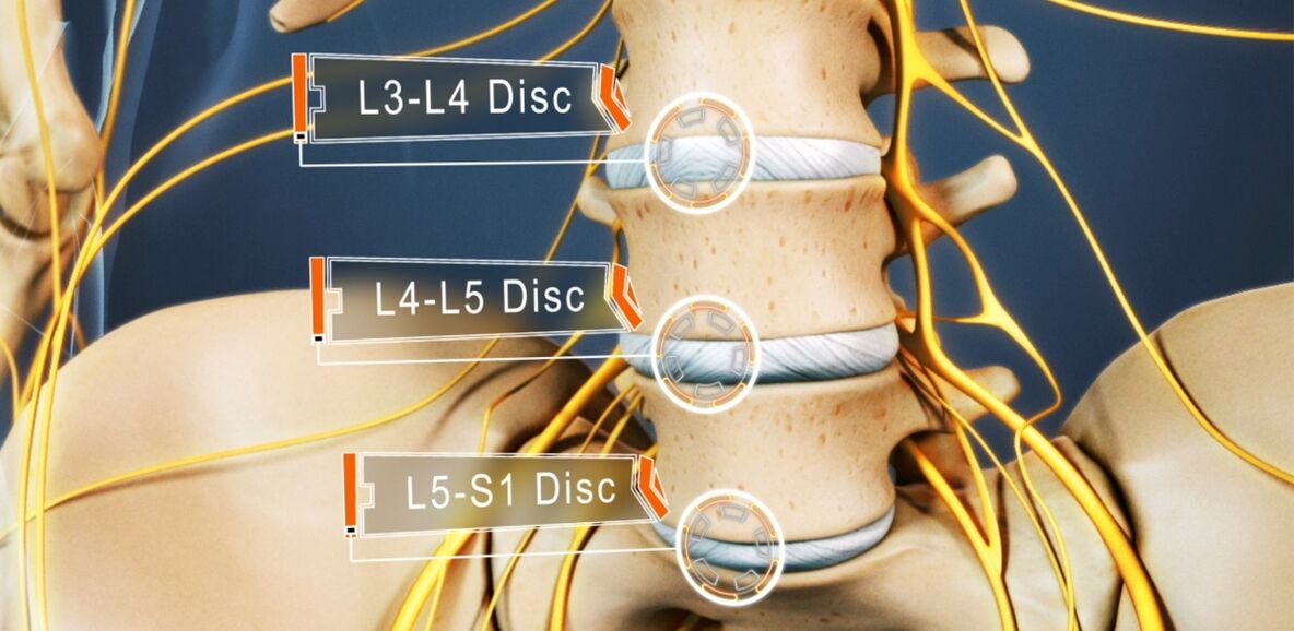
48-52% of people suffer from osteochondrosis. And osteochondrosis of the lumbar spine is the most common. The disease can affect any intervertebral disc in the lumbosacral spine, several, or even all of them. Discs L5-S1, L4-L5 suffer most often, and L3-L4 are less common. The upper lumbar disc discs (L3-L2 and L2-L1) are much less commonly affected.
The prevalence of lumbar osteochondrosis is due to the fact that the greatest load falls on the lower back during any physical work, especially lifting and carrying weights, walking, running, sitting. The lumbar spine consists of 5 vertebrae that are much larger than the thoracic and cervical vertebrae. Accordingly, the intervertebral discs separating them are larger in size. Normally, the lumbar region has a slight anterior curvature (physiological lordosis). It is the last motile part of the spine and is adjacent to the fixed sacrum, so lumbosacral osteochondrosis is most commonly referred to.
If osteochondrosis used to be an age-related disease, its first manifestations can now be seen at the age of 15-19. Among those in their thirties, 1. 1% of people already suffer from severe symptoms of degenerative-dystrophic lesions of the intervertebral discs. In the elderly (from the age of 59), the clinical manifestations of the disease are already present in 82. 5%. At the same time, the incidence of pathological diseases is constantly increasing, which is due not only to the increase in the average age of the country's population, but also to changes in lifestyle that are not improving.
Reasons for development
Today, there is still no consensus on the etiology of degenerative diseases of the spine. Nevertheless, the main theory of their development is involutiv. According to him, osteochondrosis is the result of previous damage to the intervertebral disc and spinal bone structures, as well as inflammatory and other processes. The theory suggests that degenerative changes are genetically predetermined and, in fact, inevitable. Their clinical manifestations are due to various endogenous and exogenous factors, especially in the young and middle-aged.
Thus, the development of osteochondrosis of the lumbar spine is facilitated by:
- heavy physical work, especially in connection with heavy lifting;
- sedentary, sedentary lifestyle;
- any back injury, including bruises;
- overweight;
- metabolic disorders;
- violation of posture, deformity of the spine;
- flat feet and other foot pathologies;
- pregnancy, especially multiple pregnancies.

Pathogenesis
Regardless of the cause, degeneration of the intervertebral disc occurs when the intensity of the catabolic processes (cleavage and oxidation of molecules) of matrix proteins exceeds the rate of their formation. One of the key points in this process is the malnutrition of the intervertebral discs.
Because adults, like most cartilage, do not have a direct blood supply because they do not have blood vessels, their nutrient supply and metabolic products are removed by diffusion, sequential compression and relaxation of the disc. movement. The main structure that supplies power to the plate is the end plates on the top and bottom surfaces.
The endplates themselves are a double layer formed by cells of cartilage and bone tissue. Accordingly, the side of the disc is adjacent to the plate, and the bone - to the vertebral bodies. They are characterized by a sufficiently good permeability, which ensures the exchange of substances between the cells, the intercellular material of the disc and the vessels passing through the vertebral bodies. Over the years, especially due to the negative influence of external and internal factors, the structure of the endplates changes, their blood supply decreases, which leads to a decrease in the intensity of metabolism in the intervertebral disc. As a result, its ability to produce the new matrix decreases, leading to a gradual decrease in its density with age.
At the molecular level, this is accompanied by:
- reduced rate of diffusion of nutrients and metabolites;
- decreased cell viability;
- accumulation of cell lysis products and altered matrix molecules;
- decreased production of proteoglycans (high molecular weight compounds responsible for the formation of new cartilage cells and which are the main sources of chondroitin sulfate synthesis);
- collagen rack damage.
Possible consequences
As a result of continuous changes, the intervertebral disc dries out, and the nucleus pulposus loses its proper distribution of loads. As a result, the pressure inside the disc becomes uneven, causing the fibrous ring to overload and compress in several places. Because this happens with every movement of a person, the ring is regularly subjected to mechanical pressure. This leads to unfavorable changes in it.
In addition, often a decrease in the height and elasticity of the disc leads to compensatory changes in adjacent vertebral bodies. Bone outgrowths, called osteophytes, form on their surface. They tend to increase in size over time and even merge with each other, ruling out the possibility of movement of the affected PDS.
Given that malnutrition causes damage to the collagen skeleton, pressure on the nucleus pulposus at certain points disrupts the normal structure of the fibers that make up the fibrous ring. Without intervention, this will eventually lead to cracks and fractures in them. Gradually, more and more fibers of the fibrous ring at the point of application of pressure rupture, leading to its protrusion. This is especially facilitated by the increased load on the spine. And because the lumbar region absorbs the main strain during movement and any physical activity, it suffers most often.
The protrusion of the intervertebral disc without permanent rupture of the fibrous ring and its base larger than the protruding part is called the protrusion. An intervertebral hernia with a complete rupture at each site is diagnosed.
As some of the fibers in the fibrous ring are destroyed, the pressure in the plate gradually decreases, leading to a reduction in tension and the fibers themselves. This leads to a violation of its fixation and, as a result, to the pathological mobility of the affected spinal motion segment.
The vertebral motor segment (SMS) is the structural and functional unit of the spine formed by the intervertebral disc, the adjacent vertebral bodies, their faceted joints, ligaments, and the muscles associated with these bone structures.
Normal spinal function is only possible with proper functioning of the PDS.
Symptoms of osteochondrosis of the lumbar spine
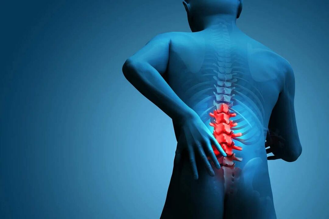
The disease may be asymptomatic for a long time and then manifest as a mild discomfort in the lumbar region, gradually intensifying. But in some cases, lumbar osteochondrosis begins acutely, immediately triggering severe pain syndrome. In most cases, signs of pathology first appear after 35 years.
Back pain is the main symptom of the disease. It can be different in nature and can be painful and dull at the same time, as well as acute, permanent, or episodic. But basically pathology, especially in the early stages of development, is characterized by alternating periods of exacerbation and remission, and both hypothermia, or the lifting of a heavy object, and unsuccessful, sudden movement can cause another deterioration in well-being.
The pain is often accompanied by numbness and tension in the back muscles. They are aggravated by physical exertion, sudden movements, heavy lifting, bending, and even coughing and sneezing.
If an anatomical structure protrudes from the spinal cord due to instability of the vertebral bodies, it leads to the development of appropriate neurological disorders. Their main manifestations are:
- shooting, severe pain radiating to the sacrum, buttocks, lower extremities, or barrier;
- hypersensitivity disorders of varying severity;
- movement restrictions, lameness;
- weakness of the muscles innervated by the trapped nerve.
In the lumbar spine, it ends at the level of 1-2 vertebrae in the spinal cord and passes into the so-called cauda equina, which is formed by the accumulation of spinal roots. In addition, each is responsible not only for the innervation of the muscles, but also for some organs in the pelvis, so prolonged compression can interfere with the work of the proper organ. This can lead to impotence, infertility, gynecological diseases, hemorrhoids and other disorders.
The clinical picture of osteochondrosis of the lumbar spine, especially in the case of long course and compression of the roots of the spine, is highly dependent on the level of the lesion, i. e. , which plate has undergone degenerative-dystrophic changes.
- Defeat of the L3-L4 plate - pain in the front and inside of the thigh, lower leg and inner ankle. This results in a decrease in the sensitivity of the anterior surface of the thigh, a severity or loss of knee sprain, and a decrease in the strength of the quadriceps muscle.
- Defeat of the L4-L5 plate - pain ranges from the top of the buttocks to the outside of the thighs and legs. Less commonly, this involves the spread of pain in the back of the leg, including the 1-3 toes. Sensitivity and muscle weakness decrease in these areas. Sometimes hypotrophy and incomplete extension of the big toe develop.
- Damage to the L5-S1 cartilage - pain begins in the middle zone of the buttocks and descends to the heel along the back or back surface of the thighs and legs and can grip the outer edge of the foot, such as 4-5 toes. Decreased sensitivity in these areas of the lower extremities, the size of the gastrocnemius and gluteus maximus are often reduced, leading to their weakness. Decreased or lost Achilles and plantar reflexes are observed when the spinal cord passing through this PDS level is affected.
Plates L1-L2 and L2-L3 are rarely affected.
The pain that accompanies the disease limits a person and significantly reduces their quality of life. Because they persist for a long time and recur regularly, even if they are not constantly present, this can only affect the psycho-emotional state. As a result, more than half of the patients experience chronic emotional stress, depressive disorders, and so on.
Diagnostics
If there are signs of osteochondrosis of the lumbar spine, consult a neurologist or vertebrologist. First of all, the doctor collects a medical history, which clarifies the nature of the complaints, the characteristics of the pain, the conditions of their occurrence and reduction, the characteristics of a person's working life, and so on.
The second stage of diagnosis, which is performed as part of the first medical consultation, is a physical examination. In doing so, the doctor assesses the condition of the skin, posture, the depth of the physiological curves of the spine, the presence of curvature, and so on. It is necessary to assess the condition of the paravertebral muscles surrounding the spine, as these are often painful and excessively tense, which is the body’s reflexive reaction to inflammation and discogenic pain.
The neurologist may already suspect the presence of osteochondrosis of the lumbar spine based on data obtained during examination and interrogation of the patient. But laboratory and instrumental diagnostic methods are needed to rule out possible concomitant pathologies, as well as to confirm the diagnosis and accurately determine the extent of damage, the severity of degenerative-dystrophic changes in the intervertebral disc, and the involvement of bone structures.
Laboratory diagnostics
The various analyzes are not conclusive in the diagnosis of lumbar spine osteochondrosis. Rather, they are aimed at assessing the extent of the inflammatory process and detecting concomitant abnormalities.
Here's how to assign:
- UAC;
- OAM;
- blood test to determine sugar levels;
- blood chemistry.
Instrumental diagnostics
All patients with suspected osteochondrosis of the lumbar spine have:
- X-ray of the lumbar spine in two projections - allows the determination of the structure of bone structures, the detection of anomalies, the formation of osteophytes, changes in faceted joints, etc. ;
- CT - allows the detection of changes in bone structures in earlier stages of development, such as the identification of indirect signs of X-rays and osteochondrosis;
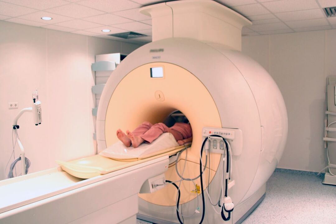
- MRI is the best method for diagnosing abnormalities in cartilage formation and other soft tissue structures, allowing the detection of the smallest lesions in the intervertebral discs, ligaments, blood vessels, and spinal cord, and an accurate assessment of their severity and potential risks.
In addition, it may be recommended that:
- densitometry - a method of determining bone density that allows the diagnosis of osteoporosis, which is particularly common in the elderly;
- myelography - allows the assessment of the condition of the CSF pathways in the spinal cord and the extent of damage to the protruding plate, which is especially important in the presence of an already formed intervertebral hernia of the lumbar spine.
Treatment of lumbar osteochondrosis
In the diagnosis of osteochondrosis, conservative therapy is usually prescribed to all patients, provided there is no pronounced and progressive neurological deficiency. But his character is strictly chosen individually.
Because the disease is chronic and the regenerative capacity of the discs is extremely limited, especially in severe degenerative-dystrophic lesions, the main goal of therapy is to stop further progression and eliminate symptoms that disturb the patient. Therefore, treatment is always complex and involves:
- drug therapy;
- manual therapy;
- physiotherapy;
- practice therapy.
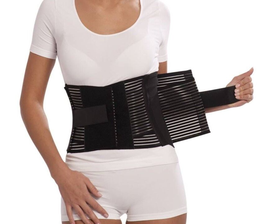
During the acute period, patients have to limit physical activity or maintain bed rest for up to 1-2 days. This helps relax the muscles and reduces the pressure inside the disc. If you need to sit, walk, or do physical work for extended periods of time, wear a stabilizing lumbar corset.
After the end of the acute period and on the contrary during the remission of the disease, it is important to move as much as possible, but carefully and to avoid increased stress in the lower back. Patients need to master the skills of sitting correctly, lifting objects off the floor, and transporting heavy loads, all of which affect the course of the pathology. It is important to avoid tilting and sudden movements, lift something off the floor or low surfaces after bending the knee, and do not bend it down. Only sit with your back straight in a chair that supports your back well. In addition, during sitting work, it is important to take regular breaks for a short workout. It is important to avoid falls, jumps, fast runs and hypothermia.
In the case of osteochondrosis, it is important to keep the body weight within optimal limits, and in the case of obesity, diet and exercise appropriate to the patient's condition are recommended, as being overweight causes increased load on the lower back and progresses more quickly. the plates.
On average, conservative therapy is usually planned for 1 to 3 months, although this can take longer. But even after completing the main course prescribed by your doctor, you should continue to take many medications, exercise therapy, and follow lifestyle recommendations.
Medical therapy
The main components of drug therapy are drugs individually selected from the NSAID group. In selecting them, the physician considers not only the severity of the pain syndrome and the course of the inflammatory process, but also the nature of concomitant diseases, especially the digestive system, as long-term NSAIDs can adversely affect the condition of the disease. mucous membranes and causes exacerbation of various pathologies of the gastrointestinal tract.
NSAIDs are required for acute lower back pain and immediately after their occurrence. Preferably within 1-2 days. Depending on the severity of the patient's condition, they may also be administered intramuscularly, rectally, suppositories, orally. The duration of the recording may not exceed 2 weeks. In the future, they will take an individually selected medication, but try to avoid frequent use.
Recently, drugs containing selective inhibitors of cyclooxygenase-2 have become more preferred.
In addition, patients are prescribed the following groups of medicines:
- muscle relaxants - help relax excessively tense muscles and thus reduce back pain;
- chondroprotectors - improves the course of metabolic processes in the intervertebral disc (especially effective when they begin at the earliest stage of the development of lumbar osteochondrosis);
- B vitamins - help improve nerve conduction;
- antidepressants and anxiolytics - used for long-term osteochondrosis that has led to depression, chronic fatigue, and other psychiatric disorders.
Therapeutic blockades are performed for very severe pain, especially of neurological origin. These include the introduction of anesthetics in combination with corticosteroids at points near the compressed nerve, leading to rapid resolution of pain. However, the procedure should only be performed by specially trained healthcare professionals in a healthcare facility as there is a risk of complications.
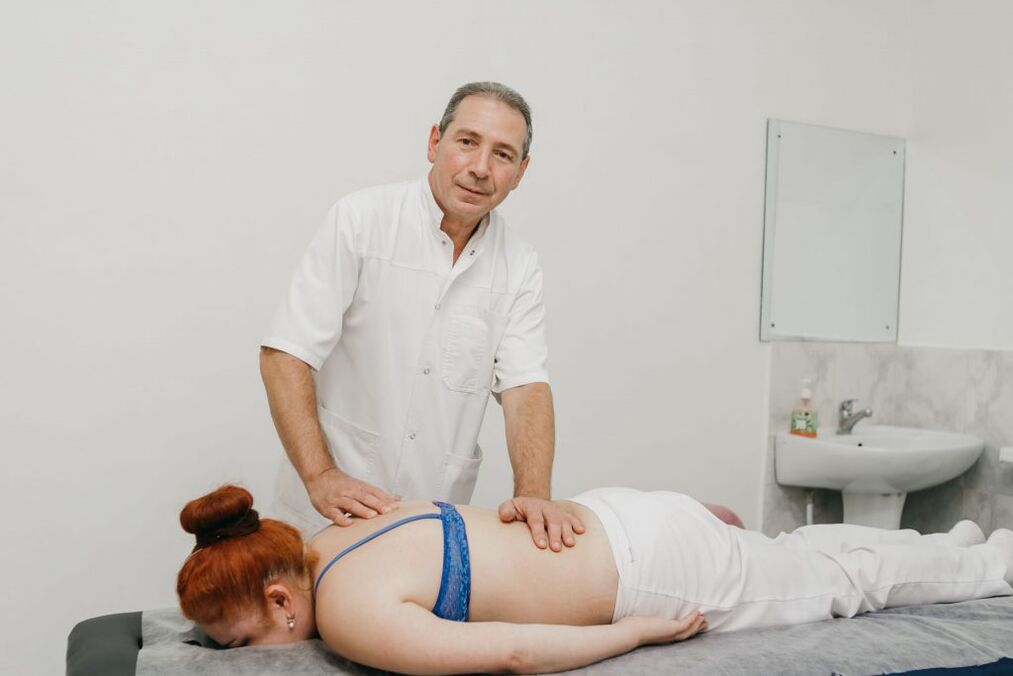
Manual therapy
Manual therapy not only improves the quality of blood circulation in the affected area, but also significantly reduces the severity and duration of pain in osteochondrosis. It effectively relieves muscle tension and allows the removal of functional blocks, which significantly increases mobility in the affected SMS.
Also, with well-conducted manual therapy, it is possible not only to increase the distance between the vertebrae and return them to their anatomically correct position, but also to release the compressed nerve roots. As a result, the pain disappears quickly and the neurological disorders disappear. It reduces the likelihood of complications and abnormalities in the work of internal organs.
Other positive features of manual therapy include improving mood, strengthening immunity, activating the body’s natural recovery mechanisms, and increasing effectiveness. In general, there is a noticeable improvement in well-being after the 1st session, and the impact will become even more pronounced in the future. The course usually consists of 8-15 sessions and it is important to complete it by the end, even if your well-being is fully normalized.
Physiotherapy
After the alleviation of acute inflammation, physiotherapy procedures are needed that not only reduce pain but also improve microcirculation, nutrition, and the course of reparative processes in degenerative-dystrophic lesions. Most often, patients are prescribed:
- introduction of electrophoresis drugs;
- electrical neuromiostimulation;
- ultrasound therapy;
- laser therapy;
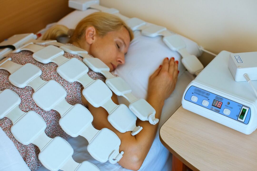
- magnetotherapy;
- UHF.
Which specific methods of physiotherapy provide the best effect, the frequency of their implementation, the duration of the course and the possibility of combining it with other types of exposure are determined individually for each patient.
Traction therapy gives very good results in lumbar spine osteochondrosis. As a result, the distance between the vertebral bodies can be increased, which immediately reduces the load on the affected cartilage discs. After the session, the patient should wear an orthopedic corset to consolidate the results.
practice therapy
After the resolution of the acute pain, the treatment program is necessarily supplemented with gymnastics. Its main tasks are to stretch the spine and relax the spasmodic muscles of the lower back. In addition, therapeutic exercises help strengthen the muscular corset, provide reliable support for the spine, and improve posture. This inevitably activates blood circulation and improves metabolic processes, which has a beneficial effect on the nutrition of the cartilage discs.
A series of exercises is selected individually for each patient, according to the degree of degenerative-dystrophic changes, the patient's level of physical development, the nature of the concomitant disorders, age, and other factors. Initially, it is recommended to study under the guidance of an experienced movement therapy instructor.
Patients with degenerative changes in the spine are recommended to visit the pelvis 2-3 times a week, as swimming instruction minimizes spinal strain but allows for effective strengthening of the back muscles.
Thus, osteochondrosis of the lumbar spine is one of the most common diseases. At the same time, it can deprive a person of their ability to work for a long time and can even lead to disability due to complications. Therefore, it is important not to ignore the first symptoms of the disease when it is easiest to treat. If you experience pain, and even more so numbness, limited mobility, and back pain, you should contact a neurologist as soon as possible, perform the necessary examination, and begin treatment. In this case, it is possible to stop the pathological process and return to normal, full life without pain and significant limitations.