


Gonarthrosis of the knee joint is the most common localization of degenerative dystrophic disease, characterized by the gradual destruction of cartilage, with subsequent changes in joint surfaces, accompanied by decreased pain and mobility.
The disease is more likely to affect women over the age of 40, especially those with varicose veins in the overweight and lower extremities.
The knee joint consists of three parts:
- medial tibiofemoral;
- lateral tibiofemoral;
- suprapatellar-femoral.
These compartments can be affected by deforming osteoarthritis (DOA) individually or in any combination. 75% of cases of gonarthrosis are damage to the medial tibiofemoral compartment (a load that is 2-3 times the body weight during exercise).
In young patients, only one joint dies more often - the right or left (right or left gonarthrosis).
Causes of DOA of the knee joint
Several factors can play a role in the development of degenerative cartilage changes at the same time:
- mechanical overload of the knee joint (certain disciplines, sports) with microtraumatization of the cartilage;
- consequences of injuries, surgical procedures (meniscectomy);
- inflammatory diseases of the knee (arthritis);
- anatomical inconsistencies in the joint surfaces (dysplasia);
- violation of statics (flat legs, curvature of the spine);
- chronic hemarthrosis (accumulation of blood in the joint cavity);
- metabolic pathology (gout, hemochromatosis, chondrocalcinosis);
- overweight;
- violation of the blood supply to the bone;
- osteodystrophy (Paget's disease);
- neurological diseases, loss of sensation in the limbs;
- endocrine disorders (acromegaly, diabetes mellitus, amenorrhea, hyperparathyroidism);
- genetic predisposition (common forms of osteoarthritis);
- a II. violation of the synthesis of type I collagen.
But in 40% of cases, it is impossible to determine the cause of the disease (primary arthrosis).
Pathogenesis of gonarthrosis
in the initial stages
In the early stages of the disease, cartilage metabolism is disrupted. The synthesis and quality of proteoglycans, the main structural unit of cartilage tissue, which is responsible for the stability of the collagen network structure, is reduced.
As a result, chondroitin sulfate, keratin, and hyaluronic acid are leached from the mesh, and structurally defective proteoglycans can no longer retain water. It is absorbed into collagen, whose swollen fibers lead to a decrease in the resistance of cartilage to stress.
Inflammatory substances accumulate in the joint cavity, causing cartilage to die even faster. Fibrosis of the joint capsule develops. Changes in the composition of the synovial fluid make it difficult to deliver nutrients to cartilage and impair the slippage of joint surfaces during movement.
Progression of pathology
In the future, the cartilage will gradually thin out, become rough, and cracks will form in its full thickness. Bone epiphyses are exposed to increased load, which causes the development of osteosclerosis and compensatory proliferation of bone tissue (osteophytes).
This reaction of the body is aimed at increasing the area of the joint surfaces and redistributing the load. But the presence of osteophytes increases discomfort, deformity, and further restricts limb mobility.
Bone fractures form in the thickness of the bone, damaging the blood vessels and leading to intraosseous hypertension. In the last stage of osteoarthritis, the joint surfaces become completely free, deformed, and limb movements are sharply restricted.
Symptoms of gonarthrosis of the knee joint
Arthrosis of the knee joint is characterized by a chronic, slowly progressive course (months, years). The clinic is gradually increasing without pronounced exacerbations. The patient does not remember exactly when the first symptoms appeared.
Clinical manifestations of gonarthrosis:
- pain. At first it is diffuse, short (walking on stairs, walking up stairs), and as osteoarthritis progresses, the pain becomes localized (the anterior and inner surfaces of the knee), increasing in intensity;
- local tactile sensitivity. Mostly on the inside of the knee, along the edge of the joint gap;
- crunch. In stage I it may not be audible, in stages II-III. accompanies every movement in the stage;
- increase in volume, deformation of the knee. As a result of the weakening of the lateral ligaments, an O-shaped configuration of the limbs develops in humans (this can be clearly seen in the photograph);
- restrictions on mobility. At first there are difficulties with bending the knee, later - with extension.
Causes of pain in the DOA:
- mechanical friction on damaged joint surfaces;
- increased intraosseous pressure, venous congestion;
- synovitis association;
- changes in periarticular tissues (stretching of the capsule, ligaments, tendons);
- thickening of the periosteum;
- phenomena of dystrophy in adjacent muscles;
- fibromyalgia;
- compression of nerve endings.
In contrast to coxarthrosis, knee DOA may show spontaneous regression of symptoms.
Clinical manifestations of gonarthrosis depending on the stage:
| Features | I set the stage | Section II | Section III |
|---|---|---|---|
| Pain | Short, more common when the knee is extended (prolonged standing, walking on stairs) | It disappears after a moderate night rest | It is stressful and disturbing at night |
| Restriction of movement | Not visible | There is a limit to expansion, slight lameness | Durable flexion-extensor contractures, lameness |
| to crack | No | You can feel it while moving | distant cracking |
| Deformation | Missing | Slight deviation of the axis of the limb at the front, muscle atrophy | Valgus or varus deformity. The joint is unstable, the atrophy of the thigh muscles |
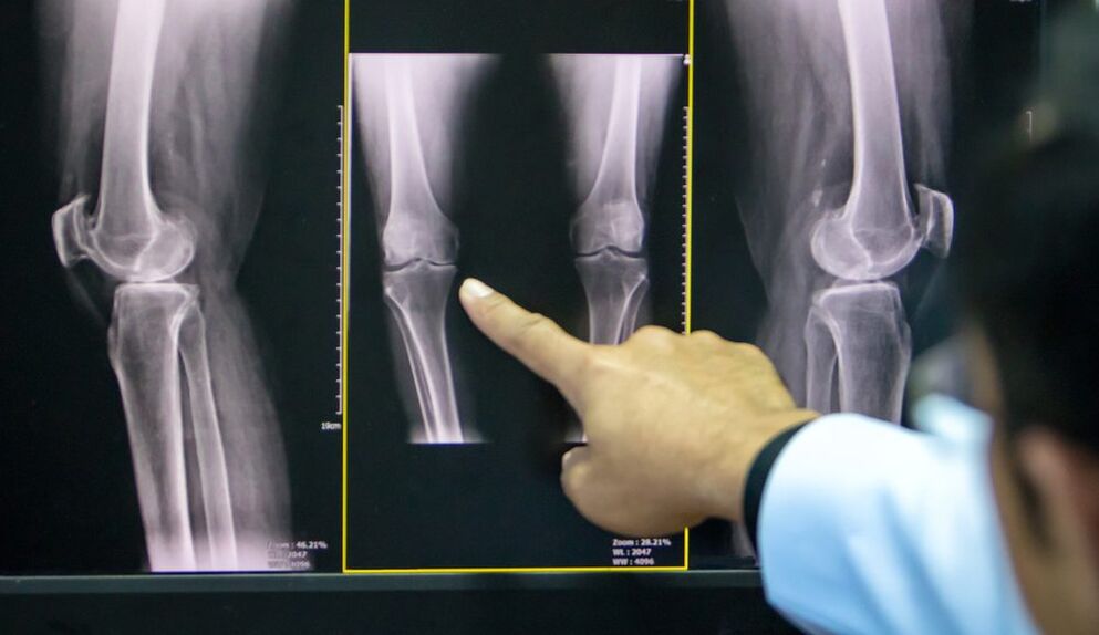
| X Ray image | Mild narrowing of the joint gap, initial signs of subchondral osteosclerosis | The joint space narrows by 50% or more, and osteophytes appear | Almost complete absence of joint space, significant deformation and sclerosis of joint surfaces, areas of subchondral bone necrosis, osteoporosis |
A common complication of knee arthrosis is secondary reactive synovitis, which is characterized by the following symptoms:
- increasing pain;
- puffiness;
- effusion into the joint cavity;
- increase in skin temperature.
Uncommon and more dangerous complications include joint blockade, osteonecrosis of the femoral condylus, subluxation of the patella, and spontaneous hemarthrosis.
Diagnosis of DOA of the knee joint
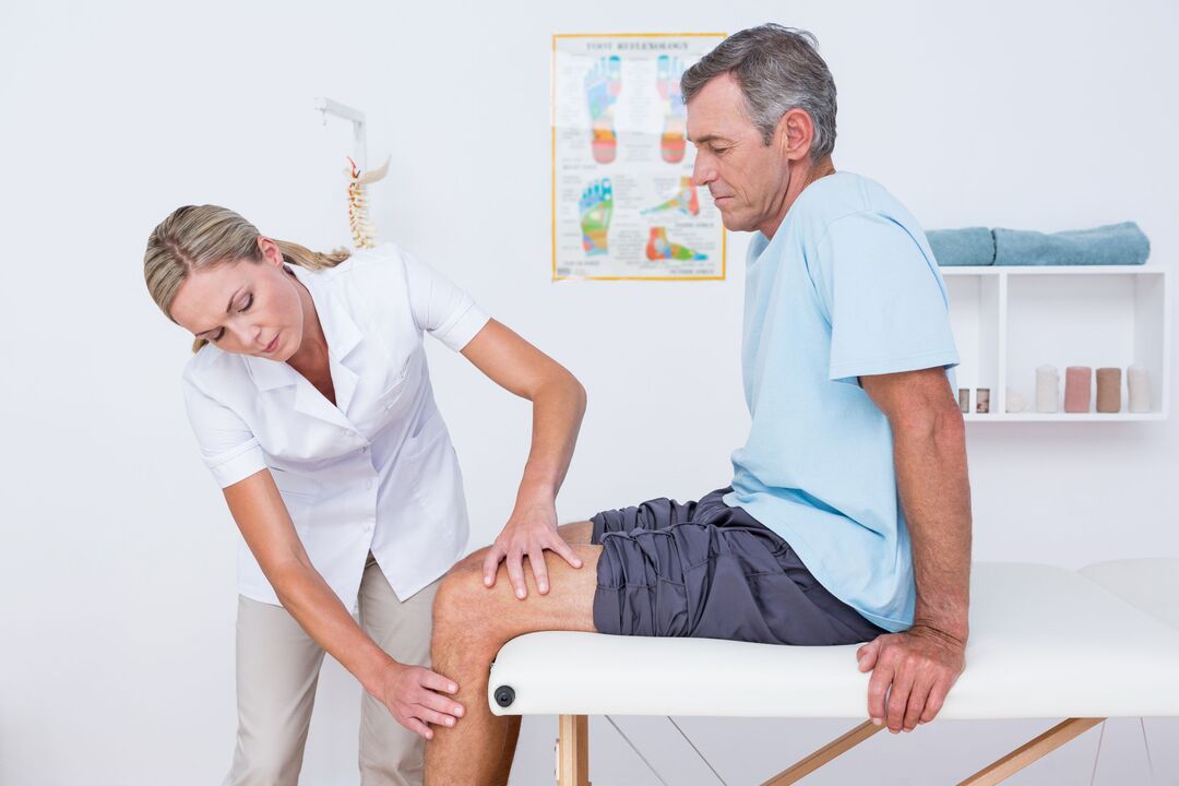
Gonarthrosis is diagnosed based on the patient's characteristic complaints, the lesions observed during the examination, and the results of further examinations.
To confirm osteoarthritis:
- radiography of the knee joint in two projections (anteroposterior and lateral): the most accessible way to confirm the diagnosis at an advanced stage of pathology;
- Ultrasound: determination of the presence of effusion in the joint, measurement of cartilage thickness;
- analysis of synovial fluid;
- diagnostic arthroscopy (visual assessment of cartilage) with biopsy;
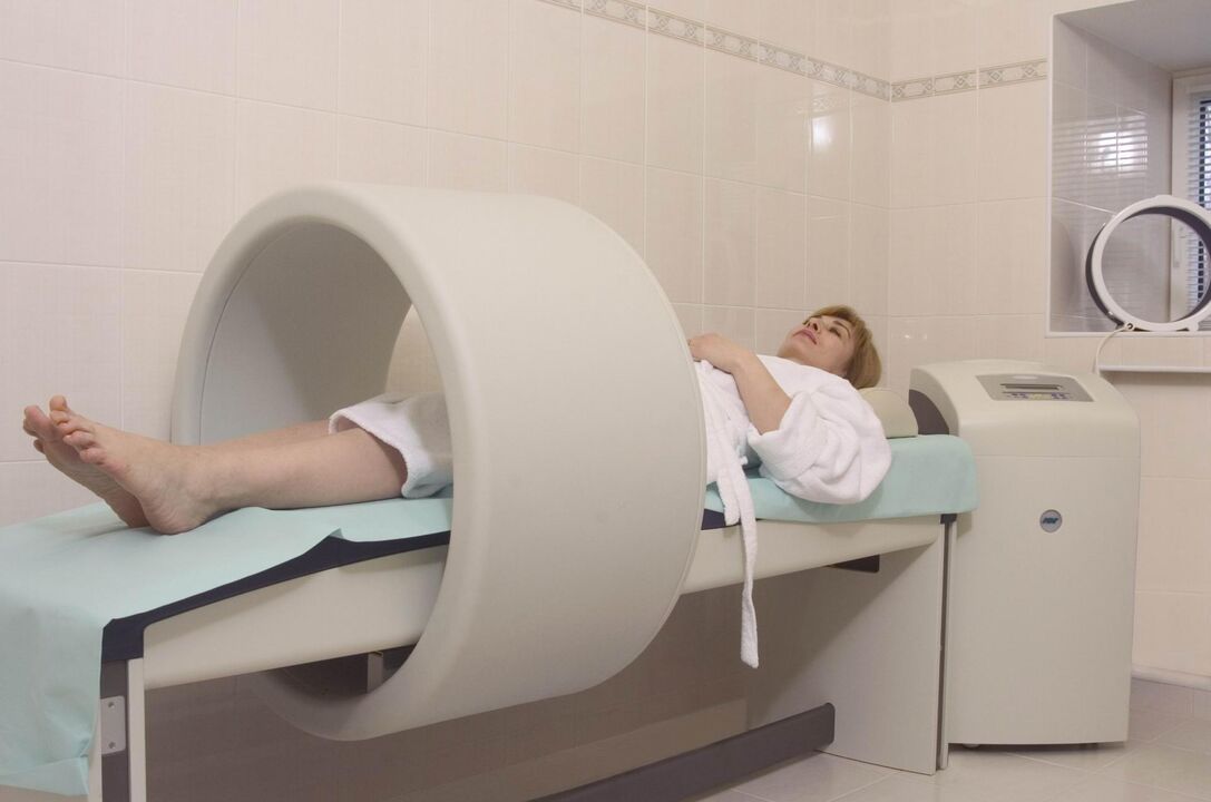
- computer and magnetic resonance imaging (CT, MRI): the best way to diagnose DOA at an early stage.
If your doctor has any doubts about the diagnosis, they may prescribe:
- scintigraphy: scanning of the joint after introduction of a radioactive isotope;
- thermography: examination of the intensity of infrared radiation (its intensity is directly proportional to the intensity of the inflammation).
Treatment of gonarthrosis of the knee joint
The treatment regimen for osteoarthritis combines several approaches: non-drug methods, drug therapy, and surgical correction. The proportion of each method is determined individually for each patient.
Non-pharmacological treatment
In the latest ESCEO (European Society for the Clinical Aspects of Osteoporosis and Osteoarthritis) guidelines for the treatment of knee inflammation, experts place particular emphasis on educating patients and making lifestyle changes.
The patient needs:
- explain what the essence of the disease, adjust to long-term treatment;
- teaching the use of aids (stick, orthosis);
- prescribes diet (for patients with a body mass index greater than 30);
- give a series of exercises to strengthen the thigh muscles and relieve the load on the knee joint;
- explain the importance of increased physical activity.
In the early stages of knee arthrosis, physiotherapy treatments give good results:
- massage;
- magnetotherapy;
- UHF therapy;
- electrophoresis;
- hydrogen sulfide baths;
- paraffinic applications;
- acupuncture.
Pharmacotherapy of gonarthrosis
The drugs used in DOA are designed to relieve pain, reduce inflammation, and slow the rate of cartilage destruction.
Symptomatic treatment:
- analgesics;
- non-steroidal anti-inflammatory drugs (NSAIDs) belonging to the group of COX-2 inhibitors in the form of tablets or suppositories;
- non-narcotic analgesics (with resistant pain syndrome).
Chondroprotectors:
- Chondroitin sulfate;
- Glucosamine sulfate.
These drugs can be taken in the form of capsules several times a year, injected intramuscularly or directly into the joint cavity.
Topical therapy consists of close and intraarticular injections of glucocorticosteroids and hyaluronic acid preparations.
DOA I – II. The use of anti-inflammatory ointments, gels and creams based on NSAIDs is an important part of complex therapy. They help the patient take oral NSAIDs, reducing the risk of damage to the digestive system.
Folk remedies
Topical application of tinctures, decoctions, extracts, and herbs is considered an adjunct to the treatment of DOA, and folk remedies are not a substitute for the therapy prescribed by a doctor.
Plants used in osteoarthritis: dandelion, ginger, Jerusalem artichoke, burdock, garlic, sea buckthorn.
Surgery
Surgery may be required at all stages of gonarthrosis, with insufficient medical intervention. Endoscopic procedures are the most common, and replacement of the endoprosthesis is recommended in the most severe cases.
Types of endoscopic surgery:
- joint review and rehabilitation: removal of inflammatory contents from the joint cavity, cartilage fragments;
- plasma or laser ablation: removal of mechanical obstructions in the joint cavity;
- cartilage surgery.
Corrective periarticular osteotomy is recommended for patients with an initial onset of axial limb deformity (up to 15-20%).
The goal of the surgery is to restore the normal configuration of the joint, distribute the load evenly on the joint surface, and remove the damaged areas. This procedure makes it possible to delay arthroplasty.
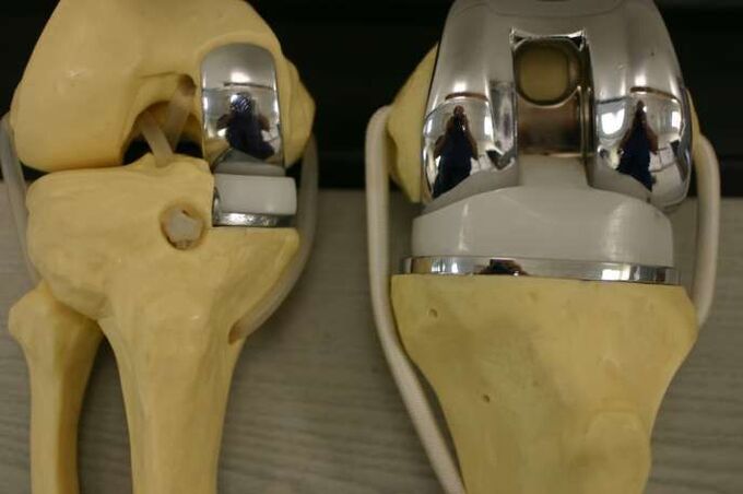
Indications for artificial replacement of the affected area (or the whole joint):
- DOA II-III grade;
- severe axial deformity of the limb;
- aseptic necrosis of the subchondral bone layer;
- persistent pain syndrome.
Contraindications to knee surgery:
- complete damage to the joint;
- unstable tape device;
- DOA as a consequence of inflammatory arthritis;
- persistent flexion contracture, severe muscle weakness.
In this case, the patient goes through arthrodesis - a comparison of the knee joint in a physiological position with the removal of the joint surfaces. This relieves pain but shortens the leg, causing secondary lesions in the opposite knee, hip and spine.
Prevention
Prevention of early cartilage degeneration should begin in childhood.
Precautions:
- prevention of scoliosis;
- flat foot correction (shoes with arch support);
- regular physical education (limits heavy sports);
- exclusion of fixed postures while working.